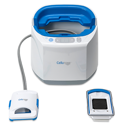
CelluTome™ Epidermal Harvesting System
The CelluTome™ System produces an array of epidermal micrografts ready for immediate transfer onto a recipient site.
Automated, uniform process
The harvesting procedure is precise and reproducible, raising uniform microdomes (photo: Full microdome formation, optimal microdome height with clear fluid encapsulated in microdome, ready to harvest into micrografts.)
Effective harvesting procedure
The CelluTome™ System consistently harvests epidermis at the dermal-epidermal junction, including basal keratinocytes. (photo: Histological microdome cross-section; 100X magnification. BK = basal layer keratinocyte; SC = stratum corneum.)
Viable Epithelium for Skin Grafting
The micrografts harvested contain undamaged tissue for grafting to a recipient site. (photo: LIVE/DEAD® stained epidermal micrograft with Calcein AM dye; green staining indicates live cells.)
Cell Proliferation
Cells grow outwards from the micrograft edge demonstrating that the cells are not damaged during acquisition. (photo: Keratinocyte outgrowth over time, cultured in EpiLife® Medium. cytoskeleton, green; plasma membrane, red; nucleus, blue; 100X magnification)